Key Skin Stem Cells and Cosmetics for Skin Regeneration

Department of Bio-Convergence Science, College of Medical Life Sciences, Konkuk University
The skin has an important function to prevent intrusion from the outside, protect the barrier, moisturize the skin, and maintain the skeleton of the skin. This requires constant normal cells to grow, typically differentiated cells such as keratinocyte and fibroblast, and stem cells are the cells that are the basis of all the cells that produce them continuously. (Webb A et al., 2006; Dahl MV. 2012).
Stem cells have two basic properties. First, self-renewal and second, pluripotency. Stem cells present on the skin have limited multifunctional differentiation, unlike embryonic stem cells, which are the source of all living cells as adult stem cells, but are the source of the skin's vitality, or new cells, and thus have great meaning of regeneration (Jones PH et al., 1995; Lenciewicz AM. 2019). Thus, since the world's first ephemeral stem cell-based cosmetics called Capture R60/80 XR were released at Christian Dior in 2007, cosmetics related to studies on stem cells have been released, with global cosmetics companies at the center, and stem cell-related products derived from keratin and dermis, and plant-derived stem cell-cell-related products have been released.
In this review paper, considering the vastness of the content, we look at recent research trends only in Figure 1 of exfoliated stem cells and dermis-derived stem cells.
1. Epidermal Stem Cells
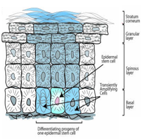
The epidermis is largely divided into four layers: basal layer, spiny layer, granular layer, and corned layer from the floor; exfoliated stem cells exist in the base layer; and they also exist in sebaceous gland, bulge region of hair follicles. Epidermal stem cells exist in microenvironmental structure called niche, and, like other stem cells, proceed self-renewal, and divide and maintain the same two daughter cells. Epidermal stem cells undergo asymmetric division to produce keratin-forming cells, producing one daughter cell identical to the stem cells, while producing another transient amplifying cell that leaves microenvironmental tissue and continues to divide, which eventually leads to differentiation for the function of skin barriers, and is eliminated from the stratum corneum after about 28 days (called desquamation).
In order to separate exfoliating stem cells from normal exfoliating cells, it was essential to find a biomarker that was uniquely expressed in exfoliated stem cells (Kretzchmar K et al., 2014). α6 integral was found to be a specific biomarker of Epidermal stem cells, and the biochemical characteristics of highly expressed epidermal stem cells showed that a6 integrin was found to have high colony forming capacity and high regeneration capacity levels when normally quiescence is or activated.(Jones PH et al., 1993; Li Aet al., 1998). The epidermal stem cells expressing β1 integrals showed rapid attachment to Type IV collagen and were also found to have high proliferative effects (Jones PH et al., 1995). CD133 (Prominin) (Yu et al., 2002) and Delta1Notch ligand (Lowell et al., 2000) were also found to be specific biomarkers of epidermal stem cells. The p63 homologue of p53 was also identified as a specific biomarker of typical epidermal stem cells (Pellegrini G et al.,2001). In addition, epidermal stem cells were also characterized by low-level expression of CD71 (Li A ethereal, 1998), the gap junction protein conexin43 (Chen Z et al., 2006), and Desmoglein3 (Wan Heet ., 2003).
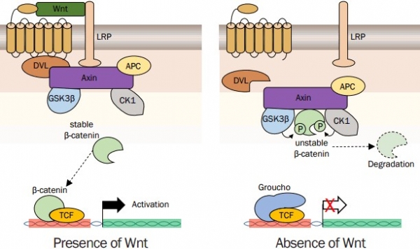
This discovery of specific biomarkers of epidermal stem cells made separation relatively easy, so studies were also conducted on the signal transmission network of epidermal stem cells. Generally, epidermal stem cells remain in a recessed state, and are strictly controlled. A study on signal transmission of the hair follicle bulge stem cell, mainly in the hair follicle, has been established, controlling cell function using sonic hedgehog (Shh) and Wnt signaling, especially the Wnt/β-catenin signal (EckertLt; Velt 2013). Wnt is a glycoprotein that binds to the Frizzled receptor. When the Wnt signal is present, the Disheveled(DVL) protein binds to Axin to deactivate GSK3β, which stabilizes --catenin and moves to the nucleus because it is not phosphatized, and causes the Wnt gene to interact with TCF1 transcription factor. On the other hand, in the absence of a Wnt signal, β-catenin is phosphatized by GSK3β and CK1 proteins, which undergoes ubiquitination and eventually decomposes. Wnt signaling plays a very important role in maintaining follicular stem cells, and inhibiting Wnt signal transmission will prevent differentiation between exfoliating cells and sebum cells (VeltriA et al., 2018; Lee JH 2017).
Sonic hedgehog(Shh) acts as another important regulator of the hair follicle bulge stem cells in the hair follicle. Sonic hedgehog(Shh) is a ligand that binds to Patched Ptc, a membrane-spanning receiver. In the recessed state, Ptc is suppressed by combining with Smoothened Smo, which is transmembrane protein. When Shh joins Ptc, Smo is free and phosphatized. The Ci protein then enters the nucleus, leading to the manifestation of the shh target genes. If you suppress the Shh signal, your hair will stop growing. (Abe Y et al., 2017)
In addition, the Notch and EGFR(epidermal growth factor receptor) signal transmission also play a very important role, with the Notch signal having high activity in the base layer of the epidermis and leading to the differentiation of keratin-forming cells when cells leave the base layer (Eckert RL et al., 2013).
Extracellular matrix (ECM) also affects the stem cell varnish of the skin. In the epidermis layer, ECM is mainly present in the basal membrane, which morphologically separates the epidermis layer from the dermis layer, which interacts with the basal membrane, and when lost, the epidermal stem cells in the recessed state divide and eventually undergo differentiation to function as a skin barrier. Destroying ECM components adversely affects the form or constancy of keratin-forming stem cells. Therefore, ECM plays a very important role in maintaining keratin-forming stem cells, appropriate migration and controlling the differentiation of keratin-forming cells (Chermnikh E et al., 2018).
In addition, it has been determined that epiderma stem cells are essential to keeping the epidermal layer normal and can also effectively contribute to the role of skin barriers, moisturizing and wound healing and burns (Li A et al., 2004; Zouboulis CC et al., 2008;Fujiwara He et al., 2018; Yang Ret al., 2019).
2. Dermal Stem Cells
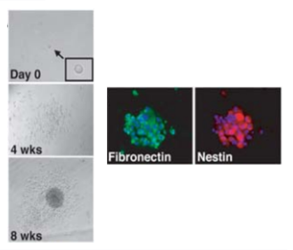
The dermis-derived stem cells are generically mesodermal lineage, are found in the dermis area and hair papillae, and have the ability to differentiate into fibroblasts and pericyte. The first dermis-derived stem cells found were separated from the neonatal human foreskin tissue and were named skin-derived precursors, SKPs. Human SKPs grew into Sphere under the conditions of mitogens fibroblast growth factor 2 and epidermal grout factor and produced certain biomarkers such as nestin, fibronectin, and vimentin (Toma JG et al., 2005).
These SKPs are resistant to UVB, and after UVB irradiation, unlike fibroblasts, which are controls, SKPs formed a cluster of globules, reduced the high expression and apoptosis levels of the proliferative indicator Ki67, and low SA-βgal, an indicator of DNA damage and aging. In addition, the expression of Bcl-2 and Nrf2 was statistically significantly high in SKPs, while the expression of Bax and Bak, cell apoptosis biomarkers, was observed low (Zhong J et al., 2016).
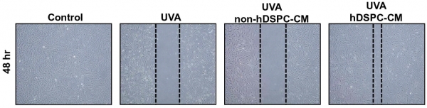
Later studies of human dermal stem cells/progenitor cells, hDSPCs were discovered in another study group and confirmed that SOX2, NANOG and S100B were expressed as specific biomarkers (Shim JH et al.,2013a; Shim JH et al., 2013b). An anti-aging study was conducted on these dermis-derived stem cells, and a study of culture badges containing ingredients secreted from dermis-derived stem cells found that various growth factors such as bFGF, IGFBP-1, IGFBP-2, HGF, VEGF and IGF-1 were expressed. As a result of treating this culture medium to fibroblasts irradiated by ultraviolet light, the increase in expression of MMP1 was significantly reduced by ultraviolet light, conversely, the expression of TIMP1, the inhibitor of the ultraviolet rays of collagen types I, IV, V and MMP1. The hDSPCs culture medium also facilitated the movement of fibrous cells damaged by ultraviolet light, and significantly reduced the effect of apoptosis by ultraviolet rays (Shim JH et al., 2013c). In other words, hDSPCs have utilized various growth factors that are secreted to minimize fibroblast damage caused by ultraviolet rays.
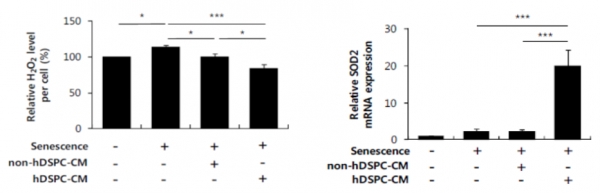
Meanwhile, the hDSPCs culture medium also showed positive results for fibrous cells suffering from cellular senses. In general, fibroblasts undergoing endogenic aging have increased the expression of aging biomarkers beta galactosidase and p53 and p21 and have been significantly reduced by hDSPCs culture badges. The hDSPCs culture medium has restored the expression of type 1 and type 3 collagen in fibroblasts suffering from endogenic aging and has shown significant reduction in the expression of MMP1 (Jung JY et al., 2015). In addition, the hDSPCs culture medium increased the expression of superoxide desmutase 2, showing the result of reducing the production of free radicals.
In conclusion, stem cells derived from dermis have been shown to be effective in improving photo-aging and endogenic aging, thus contributing to the anti-aging effect. However, unlike epidermal stem cells, the representative biomarkers have not been excavated well and research has not progressed much, so there is a slight lack of signal transmission mechanisms.
3. Application of stem cell research to cosmetics
As mentioned in the introduction, research on human skin stem cells continues to be conducted on three aspects: 1) using plant stem cells to regenerate skin stem cells based on research on human skin stem cells. 2) Using plant stem cells for safety reasons, skin regeneration is carried out as a genetic material using animal materials that are cultured in small-3) human skin stem cells. Among them, 1) and 2) have the advantage of utilizing native plants in Korea in accordance with the Nagoya Protocol.
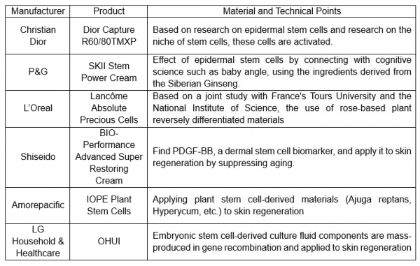
Among them, cultures or extracts, including plant growth factors, which can be obtained by cultivating growth points such as seeds, stems, and roots, are used as raw materials for cosmetics, especially plant peptide components, which are secreted by callus (referring to bloated tissue by inducing cell division when injured in a plant) in the process of differentiation into stem, leaf, flower, and various amino acids, and other amino acids. In Europe, where relatively high-tech technologies are quickly reflected in cosmetics, applications have been made around plant stem cell materials for safety reasons. The following are the first examples of stem cell-related cosmetics released by global cosmetics companies at home and abroad.
Research and development on skin stem cells is expected to continue steadily as stem cells imply skin anti-aging and regeneration in the global cosmetics market. However, it is necessary to check whether the guidelines of the Ministry of Food and Drug Safety (FDA) have been implemented well, as stem cell-related cosmetics are continuously being released based on the culture fluid or extracts of stem cells from plants or animals. According to the Ministry of Food and Drug Safety, the use of the term "stem cell cosmetics" constitutes false and exaggerated advertising, and the revision of the Regulations on the Designation of Cosmetics Materials.
Extracting a dragon is as follows.
1) 463 groups of human cells, tissues, culture fluid, asbestos, etc. have been newly designated as prohibited ingredients for cosmetics manufacturing.
2) 51 groups of ingredients, including hydrogen peroxide, that can be used within a certain range when manufacturing cosmetics.
3)In order to add salts, rename components, and add other names, 22 non-combination components, including morpholine and its salts, and 23 combination limit components, including glutarals, have been changed. However, human cell and tissue culture fluid was allowed to be used as a raw material for cosmetics only when safety was secured through donor eligibility tests, genetic toxicity tests, and skin irritation tests.
In addition, after the implementation of stem cell culture fluid cosmetics, the KFDA said, 1) False indications and advertisements containing stem cells, etc. 2) Indications and advertisements that may be mistaken for medicines
3) Indications and advertisements that advocate functionality without screening functional cosmetics 4) Management supervision was also strengthened for unfair labeling and advertising prohibited by the Cosmetics Act, such as skin regeneration and cell regeneration. For example, if you intend to develop functional cosmetics that improve hair loss symptoms by utilizing human-derived stem cell culture fluid, you should submit data in accordance with the Regulations on Functional Cosmetics Review (Article 4-Submitted Data of the Ministry of Food and Drug Safety) to be reviewed as functional cosmetics. In particular, if the government intends to apply human cell and tissue culture fluids such as "human-derived stem cell culture fluid" as raw materials for cosmetics, it should submit separate data proving that they meet the safety standards of human cell and tissue culture fluid in accordance with the Regulations on Cosmetics Safety Standards (appendix 3).
Cosmetics are rapidly and technologically upgraded as research from a skin science perspective becomes more active in line with the rapid progress of the latest bio-technology. However, although it does not require the level of medicine, if the materials that are intended to be applied as cosmetics were secured from animals rather than plants, it will be recognized by customers as a product that can be re-purchased only when research that can be effectively transmitted to the skin through safety or innovative formulation methods is conducted together.